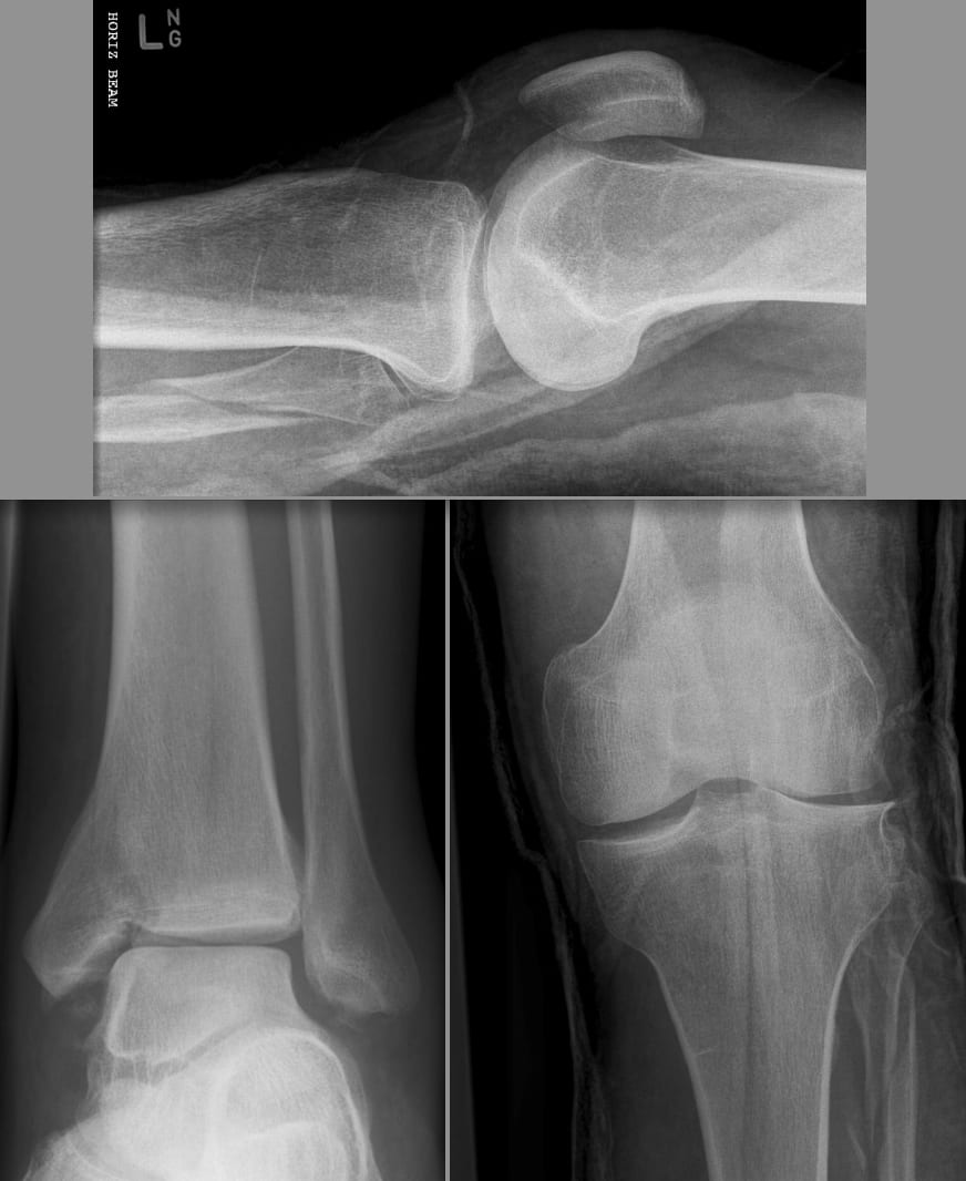
Patients generally do not report pain near the proximal fibula, so physical examination such as palpation along the fibula is effective for differentiating a Maisonneuve fracture from an isolated syndesmotic injury. Diagnosis ĭiagnosing a Maisonneuve fracture requires a combination of medical history, physical examination, and radiographic imaging. Slight or high degrees of plantarflexion prior to supination-external rotation of the foot have been identified in patients with proximal fibular fractures.

Īlthough most Maisonneuve cases report a pronation-external rotation mechanism of injury, clinical studies have recorded instances of supination-external rotation being the mechanism of injury. Diastasis of the lateral malleolus may also occur, in which it is posterolaterally displaced from the tibia. In cases where the anterior aspect of the tibiofibular syndesmosis can resist mechanical stress, only an oblique fracture of the lateral malleolus is produced. The force results in a spiral, sometimes an oblique, fracture at the neck of the proximal fibula.Rotative energy is transferred upwards along the interosseous membrane, damaging it in the process.The ankle mortise is subjected to excessive torque, rupturing the syndesmotic ligaments and anteromedial ankle joint capsule.Forceful, external rotation of the ankle joint results in the tearing of the deep deltoid ligament and/or an avulsion fracture of the medial malleolus.The following are described as subsequent events that result in a Maisonneuve fracture: The Maisonneuve fracture generally follows a specific pattern of injury. Pathophysiology įracture of the lateral malleolus seen on X-ray scan (left ankle) This leaves the ankle joint in a state of chronic pronation, characterised by a protrusion of the medial malleolus into the subcutaneous tissue. If a Maisonneuve fracture is left untreated, instability of the tibiotalar joint and deltoid ligament can cause a valgus deformity of the ankle. A long-term effect of this is painful ankle osteoarthritis due to the direct contact between the tibia and talus. Īs the syndesmotic ligaments are responsible for stabilising the ankle mortise and tibiotalar joint, disruption to this syndesmosis can cause a reduction of the space between the distal tibia, fibula, and talus. Damage to the deltoid ligament or interosseous membrane can cause haemorrhaging around the surrounding tissues, resulting in a localised oedema.

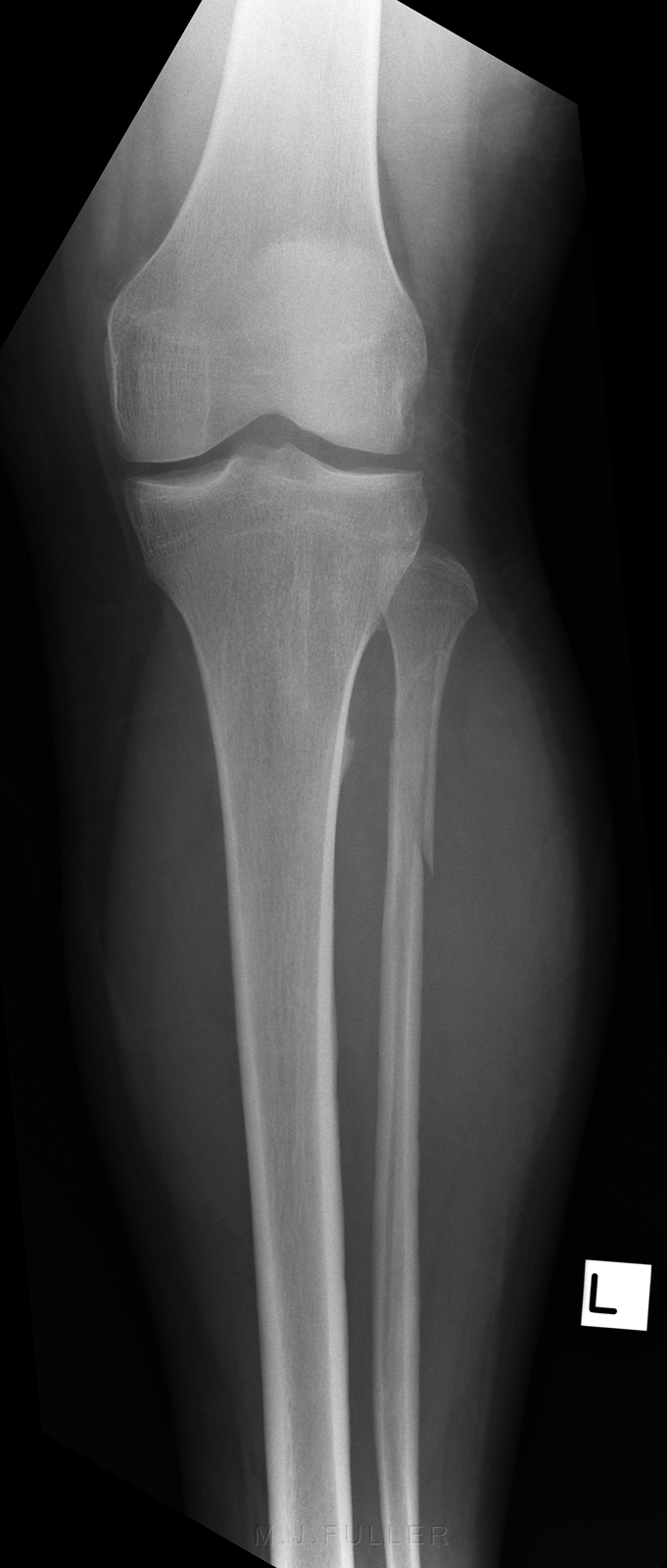
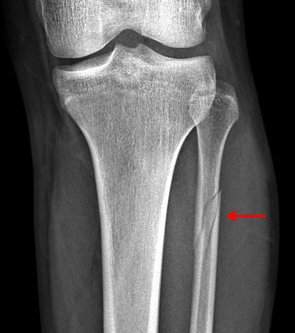
Pain may also be felt around the medial and lateral aspects of the ankle, and more rarely around the superior (or proximal) tibiofibular joint. Additionally, there is a reduced range of motion of the foot and an inability to weight-bear due to ankle pain. More specifically, as a pronation-external rotation injury, pain during external rotation of the ankle joint is expected. įracture of the medial malleolus seen on X-ray scan (left ankle)Ĭommon symptoms of a Maisonneuve fracture are pain, swelling, tenderness, and bruising around the ankle joint and inferior (or distal) tibiofibular joint. The fracture is named after the surgeon Jules Germain François Maisonneuve. The Maisonneuve fracture is similar to the Galeazzi fracture in the sense that there is an important ligamentous disruption in association with the fracture. It is also classified as a Type C ankle fracture according to the Danis-Weber classification system. Due to this, the Maisonneuve fracture is described as a pronation- external rotation injury according to the Lauge-Hansen classification system. The Maisonneuve fracture is typically a result of excessive, external rotative force being applied to the deltoid and syndesmotic ligaments. This type of injury can be difficult to detect. There is an associated fracture of the medial malleolus or rupture of the deep deltoid ligament of the ankle. The Maisonneuve fracture is a spiral fracture of the proximal third of the fibula associated with a tear of the distal tibiofibular syndesmosis and the interosseous membrane. Isolated tibiofibular syndesmosis injury, isolated fibula fracture Physical examination, radiography, X-ray, CT, MRI, arthroscopy Sporting injuries, falls, motor vehicle accidents Swelling around medial and lateral sides of ankle joint, pain during external rotation of foot

Radiograph showing a Maisonneuve fracture of the proximal fibula


 0 kommentar(er)
